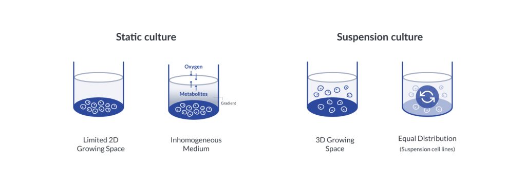
Traditional cell culture uses the monolayer culture which is also a standard procedure. However, the 2D cell culture method is not similar to the in-vivo microenvironments. 3D cell culture mimics the in-vivo microenvironments, which can improve cell growing with in vitro model.
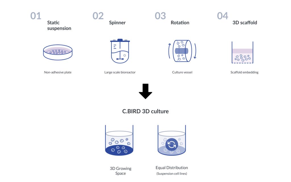
To achieve the purpose of 3D spheroid cell culture, numerous methods have been used to establish the culture environments such as suspension culture of non-adhesive plate, spinner bioreactor, rotating bioreactor, 3D scaffold etc.
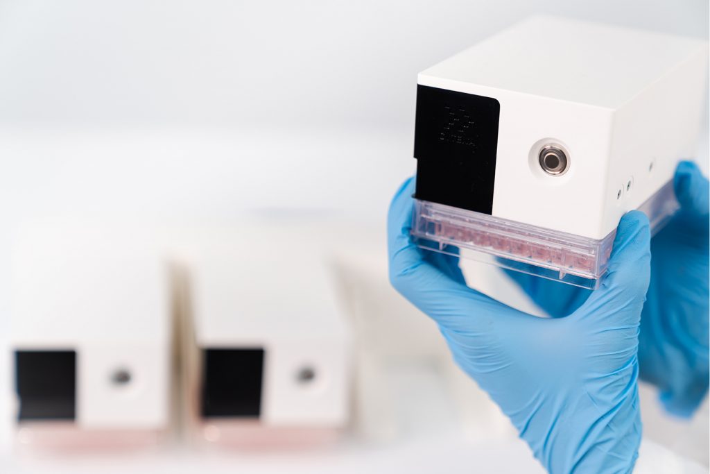
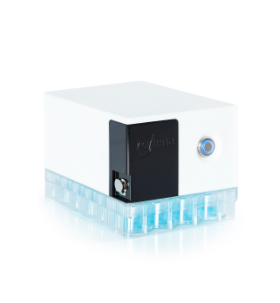
C.BIRD offers continuous mixing as a multiple pipetting system, which can keep cells in a low adhesion status. Cells will grow and aggregate into a spheroid in the standard 96/24-well plates.
Introduction
Traditional cell culture uses the monolayer culture which is also a standard procedure. However, the 2D cell culture method is not similar to the in-vivo microenvironments. 3D cell culture mimics the in-vivo microenvironments, which can improve cell growing with in vitro model.
To achieve the purpose of 3D spheroid cell culture, numerous methods have been used to establish the culture environments such as suspension culture of non-adhesive plate, spinner bioreactor, rotating bioreactor, 3D scaffold etc.
C.BIRD offers continuous mixing as a multiple pipetting system, which can keep cells in a low adhesion status. Cells will grow and aggregate into a spheroid in the standard 96/24-well plates.
Traditional methods of 3D culture
Our solution
Performance data
An Innovative 3D Cell Culture System for Tumor Spheroid
Spheroid culture, an in vitro 3D cell culture system, provides cell-cell and cell-extracellular matrix (ECM) interaction networks to retain cellular phenotype through signaling. It breaks restrictions in recapitulating in vivo microenvironments and is emerging as a powerful tool for therapeutic development, including drug screening and cancer research. In recent studies, gene and physiological expressions in 3D cell spheroids are much closer to clinical expression profiles than those seen in 2D cell monolayers.
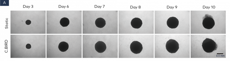
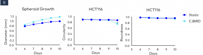
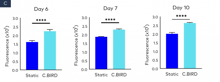
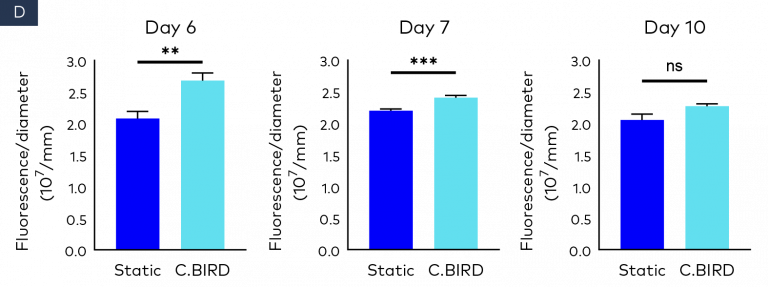
Figure 2. Comparison of spheroid growth and cell health of colon cancer HCT116 spheroids in 96-well, ultra-low attachment plates in static and C.BIRD culture. (A) Spheroid morphology of HCT116 tumor cell lines formed from 3 to 10 days was observed under microscopy. Scale bar = 0.5 mm. (B) Growth kinetics, circularity and roundness of spheroids were evaluated over a period of 10 days. The analysis was performed using ImageJ software, and data represent mean ± SEM of six replicates for each group. (C and D) Spheroid cell health was assessed using PrestoBlue Cell Viability Reagent. After 6, 7 and 10 days of spheroid culture, 20 μL of PrestoBlue Cell Viability Reagent was added to each well, which were then incubated at 37°C and 5% CO2 for an additional 3 hours before being read on a fluorescence-based microplate reader (Ex/Em ~560/590 nm). Cell viability was measured as fluorescence signals in each group with six replicates. Fluorescence signals were normalized by spheroid diameter; a higher ratio (fluorescence/diameter) indicates healthier spheroids. The significance of P values is listed as: P ≤ 0.01 (**), P ≤ 0.001 (***) and P ≤ 0.0001 (****). Data are shown as mean ± SEM.
Conclusion
In conclusion, our results have two main implications:
(1) the C.BIRD culture method improves tumor
spheroid growth in serum-free media by using a
ULA plate, and (2) the duration of spheroid viability
is prolonged by the C.BIRD mixing culture. This
means the C.BIRD culture method is a better option
to optimize the tumor spheroid 3D culture and can
improve drug screening workflows to obtain more
accurate results in viability/cytotoxicity assays.
We use cookies on our website to give you the most relevant experience by remembering your preferences and repeat visits. By clicking “Accept”, you consent to the use of ALL the cookies.
This website uses cookies to improve your experience while you navigate through the website. Out of these, the cookies that are categorized as necessary are stored on your browser as they are essential for the working of basic functionalities of the website. We also use third-party cookies that help us analyze and understand how you use this website. These cookies will be stored in your browser only with your consent. You also have the option to opt-out of these cookies. But opting out of some of these cookies may affect your browsing experience.
Necessary cookies are absolutely essential for the website to function properly. These cookies ensure basic functionalities and security features of the website, anonymously.
Cookie
Duration
Description
cookielawinfo-checbox-analytics
11 months
This cookie is set by GDPR Cookie Consent plugin. The cookie is used to store the user consent for the cookies in the category "Analytics".
cookielawinfo-checbox-functional
11 months
The cookie is set by GDPR cookie consent to record the user consent for the cookies in the category "Functional".
cookielawinfo-checbox-others
11 months
This cookie is set by GDPR Cookie Consent plugin. The cookie is used to store the user consent for the cookies in the category "Other.
cookielawinfo-checkbox-necessary
11 months
This cookie is set by GDPR Cookie Consent plugin. The cookies is used to store the user consent for the cookies in the category "Necessary".
cookielawinfo-checkbox-performance
11 months
This cookie is set by GDPR Cookie Consent plugin. The cookie is used to store the user consent for the cookies in the category "Performance".
viewed_cookie_policy
11 months
The cookie is set by the GDPR Cookie Consent plugin and is used to store whether or not user has consented to the use of cookies. It does not store any personal data.
Functional cookies help to perform certain functionalities like sharing the content of the website on social media platforms, collect feedbacks, and other third-party features.
Performance cookies are used to understand and analyze the key performance indexes of the website which helps in delivering a better user experience for the visitors.
Analytical cookies are used to understand how visitors interact with the website. These cookies help provide information on metrics the number of visitors, bounce rate, traffic source, etc.
Advertisement cookies are used to provide visitors with relevant ads and marketing campaigns. These cookies track visitors across websites and collect information to provide customized ads.